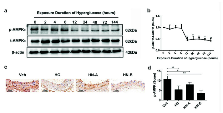
Figure 6.
Adenosine monophosphate-activated kinase (AMPK) phosphorylation was suppressed during high-glucose-induced vascular injury both in vitro and in vivo. (a,b) AMPK phosphorylation in HUVECs with different high-glucose exposure times. Data are expressed as the mean ± SEM. n = 3 for each group. * p < 0.05, ** p < 0.01 vs. normal-glucose group (the 0 h group). (c,d) Immunohistochemical staining and quantification of p-eNOS on abdominal aortic sections. Data are expressed as the mean ± SEM. n = 6 for each group. * p < 0.05, ** p < 0.01 vs. control (the Veh group).

