Figure 2.
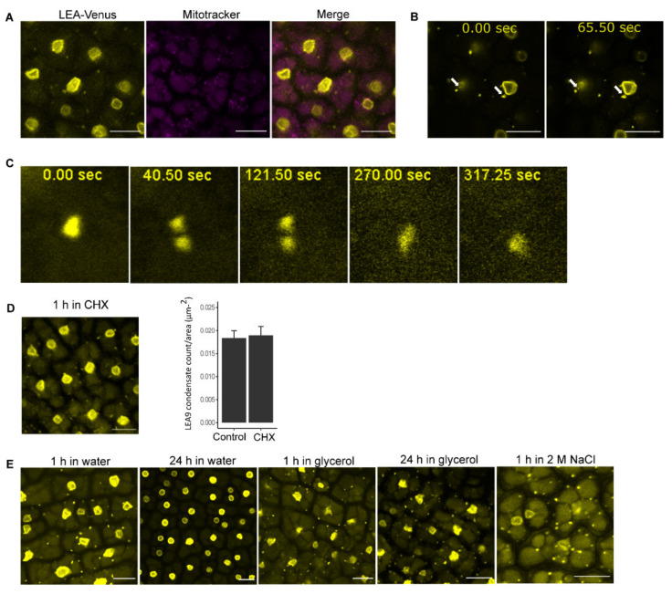
Cytoplasmic condensates of embryos of dry seeds from proLEA9::LEA9:Venus line. (A) Cytoplasmic condensates did not overlap with mitochondria by staining dissected embryos from seeds submerged in water for 1 h with Mitotracker. (B) Slight movement of cytoplasmic condensates as indicated by white arrows. (C) Division and fusion of cytoplasmic condensates. (D) Cytoplasmic condensate persistence after seeds were submerged in 1 mg/mL cycloheximide solution for 1 h. The bar plot shows counts of LEA9 condensates in seeds submerged in water or cycloheximide for 1 h (n = 9). (E) Observation of cytoplasmic condensates in dry seeds after imbibition in different conditions. Bars indicate 10 µm.

