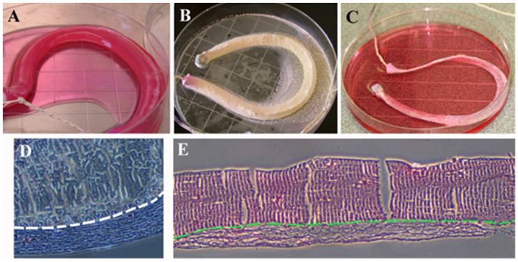
Figure 6.
Macroscopic view of lyophilization steps of the bACL and its histological features. (A) A bACL of the second generation made of native Type I collagen matrix polymerized around the braided thread scaffold before (B), its lyophilization at −80 °C and (C) its rehydration at 4 °C. Histological sections of the thread surrounded by Type I collagen were stained using the Trichrome de Masson’s (D) and the hematoxylin–eosin techniques (E). In (D,E), the structural aspect of the thread is shown above the white or the green dashed line, while the collagen seeded with ACL fibroblasts can be observed below the line (×40).

