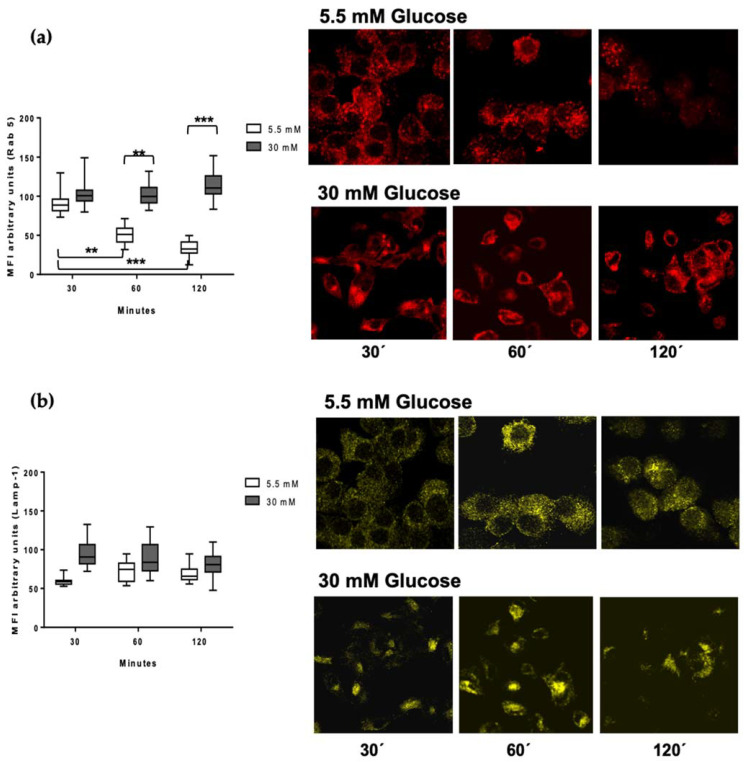
Figure 4.
Detection of Lamp-1 and Rab 5 in the processing kinetics of M. tuberculosis in 5.5 and 30 mM of glucose. MDMs were infected with M. tuberculosis previously stained with PKH67 green fluorescent for one hour and incubated at 37 °C, 5% CO2, during 30, 60, and 120 min. (a) Rab 5 (red) and (b) Lamp-1 (yellow) quantification of mean fluorescence intensity from at least 100 cells from each condition (5.5 mM and 30mM of glucose) and time point using the IMAGE J software. A representative confocal microscopy image of each condition is shown. Data were analyzed using two-way ANOVA, and the differences were considered significant when ** p < 0.001, *** p < 0.0001; from four independent experiments. Confocal image 60×, 1 zoom.

