Figure 3.
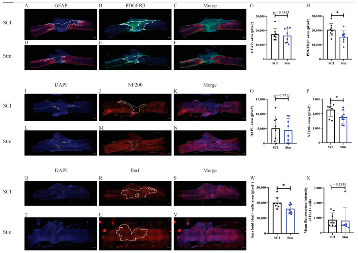
rTSMS treatment enhances tissue repair after a complete transection of the spinal cord 15 days after SCI in rats. At day 15, immunohistological analyses were performed. (A–F,I–N,Q–V) Representative pictures of sagittal spinal cord sections of (A–C,I–K,Q–S) SCI and (D–F,L–N,T–V) Stm (rTSMS treated) animals. Sections were stained with (A,D) GFAP, (B,E) PDGFRβ, (I,L,Q,T) DAPI, (J,M) NF200 and (R,U) Iba1. (G) Quantification of astrocytic-negative area (GFAP-). (H) Quantification of fibrosis-positive area (PDGFRβ+). (O) Quantification of DAPI-negative area (DAPI-). (P) Quantification of NF200-negative area (NF200-). (W) Quantification of Iba1-amyboid-positive cells’ area (Iba1+) and (X) quantification of Iba1+ mean fluorescence intensity. Scale bars are 200 µm; n = 8 animals per group. Quantifications are expressed as average ± SD. Statistical evaluations are based on the Mann–Whitney test (* = p < 0.05).

