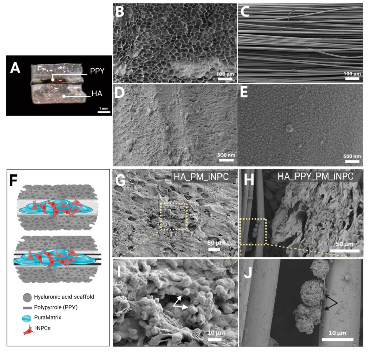
Figure 4.
Morphological analysis of iNPCs and scaffold components. (A) Macroscopic image of the HA demilune scaffold containing PPY microfibers in the lumen (hydrated state), scale bar: 1 mm. (B) SEM image detailing HA scaffold porosity. (C) SEM image showing the parallel alignment of PPY microfibers. (B,C) scale bar: 100 µm. (D) SEM image of PM on the PPY microfiber surface. (E) SEM image showing the PPY coating of the PLA microfiber surface. (B,C) scale bar: 500 nm. (F) Schematic diagram of scaffolds (HA alone and HA with PPY fibers) seeded with PM-embedded iNPCs. (G,H) SEM images of PM-embedded iNPCs seeded on HA (G) and HA-PPY (H) after three days in culture, scale bar: 50 µm. (I,J) show a higher magnification of the indicated area in (G,H) respectively, scale bar: 10 µm.

