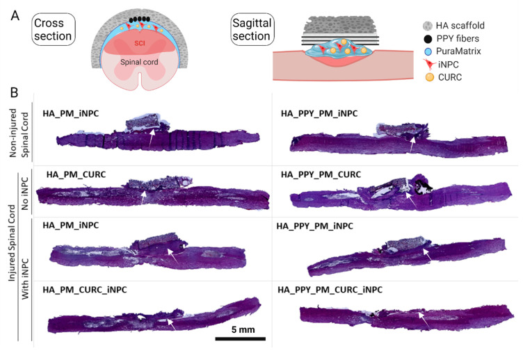
Figure 5.
Implantation of demilune scaffolds to cap the SCI. (A) Schematic diagram showing the cross and sagittal sections of the SCI and various scaffolds capping the injured area. (B) Representative images of H&E staining of sagittal spinal cord sections from one animal per group one week after injury. The white arrow indicates the formation of a fibrous-like tissue pad. Scale bar: 5 mm.

