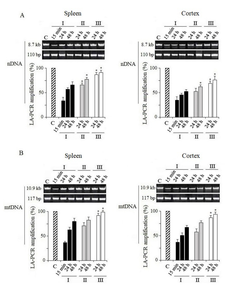
Figure 1.
Analysis of damage and repair of nuclear DNA and recovery of mitochondrial DNA. Long fragments of nDNA (8.7 kb) and mtDNA (10.9 kb) were measured. These data were normalized by the measured levels of the short fragment of nDNA (110 bp) and mtDNA (117 bp), obtained using the same DNA sample. (A) Quantitative analysis of the LA-QPCR amplicons of nDNA extracted from spleen and cerebral cortex. (B) Quantitative analysis of the LA-QPCR amplicons of mtDNA extracted from spleen and cerebral cortex. Data are presented in % to control (C). Here and in other figures: the dose of X-ray irradiation of mice was 5 Gy and MEL was administered to mice before and after irradiation as a single dose of 125 mg/kg. Electropherogram samples of synthesized amplicons are presented above the histograms. The numbers (15 min, 24 h, 48 h) above and below indicate the time after irradiation. I—mice without MEL administration; II—MEL administration before irradiation; III—MEL administration after irradiation. The data are presented as mean ± SEM of 5–6 independent experiments. Statistical significance was set at * p < 0.05.

