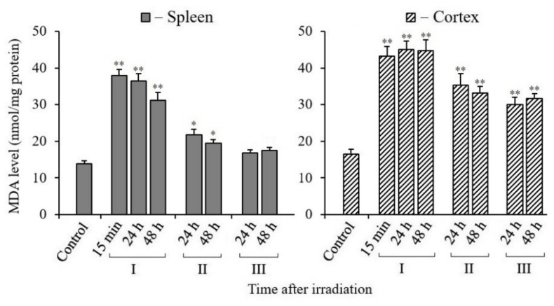
Figure 6.
Changes in the MDA content in spleen and cerebral cortex tissues of mice 15 min, 24, and 48 h after their exposure to X-rays. I—mice without MEL administration; II—MEL administration before irradiation; III—MEL administration after irradiation. The data are presented as mean ± SEM of 5–6 independent experiments. Statistical significance was set at * p < 0.05, ** p < 0.01.

