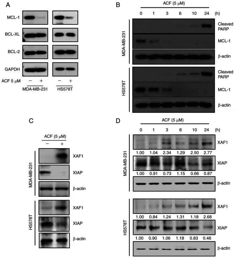
Figure 3.
ACF suppresses anti-apoptotic proteins MCL-1 and XIAP. (A) MDA-MB-231 and HS578T cells were treated with 0 or 5 µM ACF for 6 h and then western blot analysis for MCL-1, XIAP, BCL-2 and BCL-XL expression was performed. (B) MDA-MB-231 and HS578T cells were treated with 5 µM ACF and expression of cleaved PARP was assessed after 0, 1, 3, 6, 10 and 24 h using western blot analysis. (C) MDA-MB-231 and HS578T cells were treated using 0 or 5 µM ACF for 24 h and expression of XAF1 and XIAP was assessed using western blot analysis. (D) MDA-MB-231 and HS578T cells were treated using 5 µM ACF for 0, 1, 3, 6, 10 and 24 h and the expression of XAF1 and XIAP was assessed using western blot analysis. In all experiments, GAPDH or β-actin served as the loading control. ACF, acriflavine; MCL-1, myeloid cell leukemia sequence 1; BCL-2, B-cell lymphoma 2.

