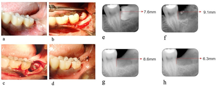
Figure 2.
Control Site. (a) Pre-operative photograph, (b) exposed impacted tooth, (c) Extraction socket on the distal aspect of the second molar, (d) primary closure of socket. (e) Pre-operative radiograph, (f) radiographs on the day of suture removal, (g) radiographs on 90th day, (h) radiographs on 180th day.

