Figure 7.
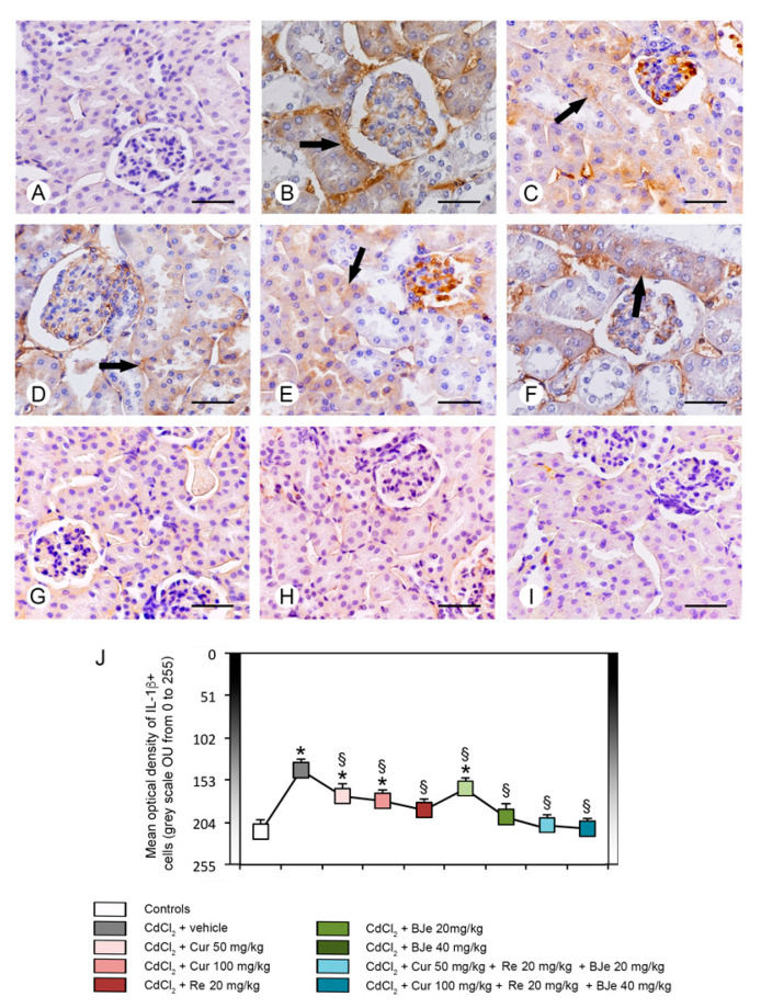
Immunohistochemical localization of IL-1β in the kidneys. (A) In all control groups, no IL-1β immunoreactivity can be demonstrated. (B) In CdCl2 plus vehicle-treated mice, nearly all tubules showed a strong IL-1β immunoreactivity (arrow). (C–E) In mice treated with CdCl2 plus both doses of Cur and with CdCl2 plus Re, a moderate IL-1β immunoreactivity was present in some tubules (arrow). (F) In CdCl2 plus BJe at the lower dose challenged mice, IL-1β immunoreactivity (arrow) was milder if compared to CdCl2 alone treated mice, but higher respect to Cur and Re. (G–I) In mice treated with CdCl2 plus BJe at 40 mg/kg and with CdCl2 plus both associations, IL-1β immunoreactivity was absent, similar to controls. (J) Morphometric results for IL-1β expression. Data are expressed in Optical Units/Unit Area (OU/UA) (from 0 = black to 255 = white). * p < 0.05 vs. control; § p < 0.05 vs. CdCl2 plus vehicle. Scale bar: 50 µm.

