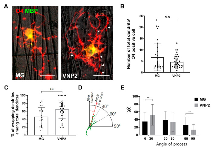
Figure 5.
Behavior of oligodendrocytes differentiated on either Matrigel or VNP2 on nanofibers coated with corresponding substrates. (A) Representative images of O4- and MBP-positive cells differentiated on nanofibers coated with Matrigel or VNP2. Arrow heads point to the processes contacting with nanofibers. (B) Quantification of total processes per an O4-positive cell differentiated on nanofibers coated with the indicated substrate. (C) Quantification of processes contacting with nanofibers per an O4-positive cell on nanofibers coated with the indicated substrate. Note that O4-positive cells differentiated on VNP2-coated nanofibers exhibit more processes that contact with nanofibers. (D) A schematic diagram illustrating how to measure the angle of processes with respect to the direction of nanofibers. (E) Quantification of process numbers according to their angle with respect to the direction of nanofibers. Scale bars: 10 µm. ** p < 0.01, Student t-test.

