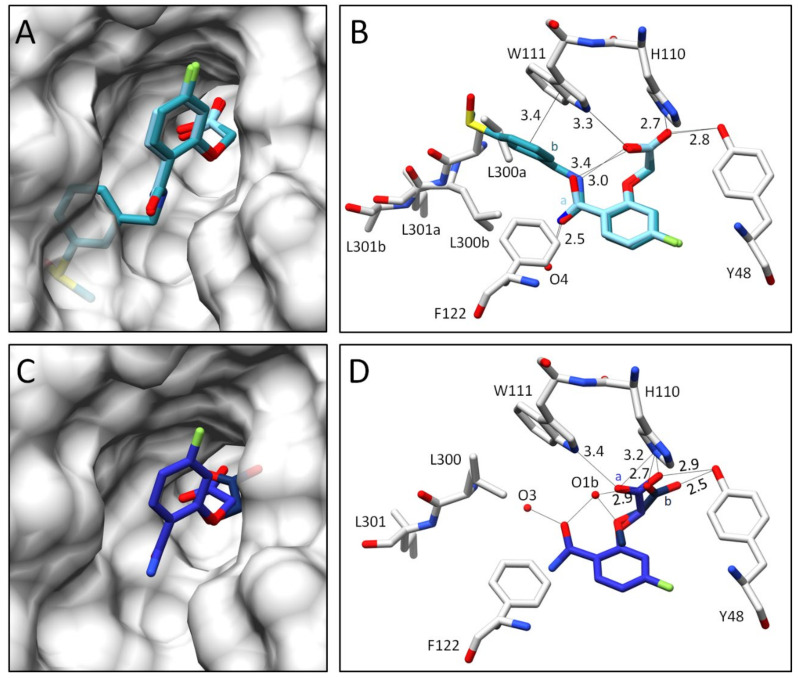
Figure 5.
Crystal structures of inhibitors 3 and 4 in complex with wild type ALR-2. Inhibitors 3 (6TUF) in light blue (A,B) and 4 (6TUC) in blue (C,D) bound to the active site of ALR-2. To distinguish the position of the conformations, b is highlighted by a slightly darker color. On the left, the protein is depicted by its transparent solvent-accessible surface (light grey) whereas on the right, the interactions are indicated as black lines. Selected residues are displayed for better orientation.

