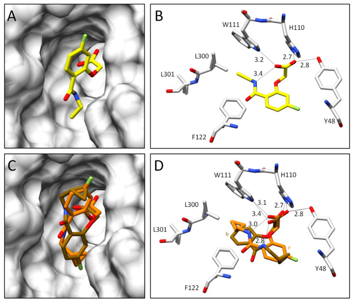
Figure 8.
Crystal structures of inhibitors 5 and 6 in complex with ALR-2. Inhibitors 5 (not deposited) in yellow (A,B) and 6 (6SYW) in orange (C,D) bound to the active site of ALR-2. To distinguish the position of the conformations, b is highlighted by a slightly darker color. On the left, the protein is depicted by its transparent solvent-accessible surface (light grey) whereas on the right, the interactions are indicated as black lines. Selected residues are displayed for better orientation. Oxygen atoms are displayed in red, nitrogen atoms in blue, sulfur atoms in yellow, and fluorine atoms in green.

