Figure 2.
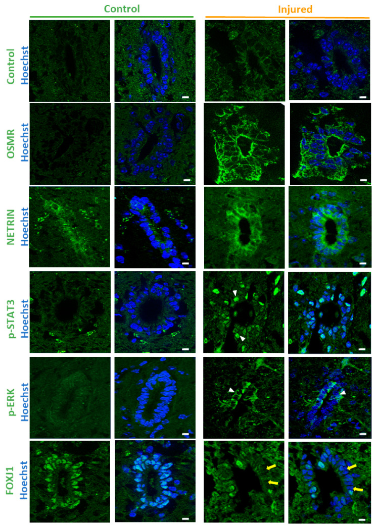
Immunofluorescence validation. Immunofluorescences performed on control and injured spinal cords for indicated proteins. Control immunofluorescence was performed using antibody against the synthetic DYKDDDDK TAG antigen. White arrowheads and yellow arrows show examples of positive and negative cells respectively. These images are representative of 2 independent experiments (n = 6 animals in total, 7–15 sections examined per animal). Scale bars = 10 µm.

