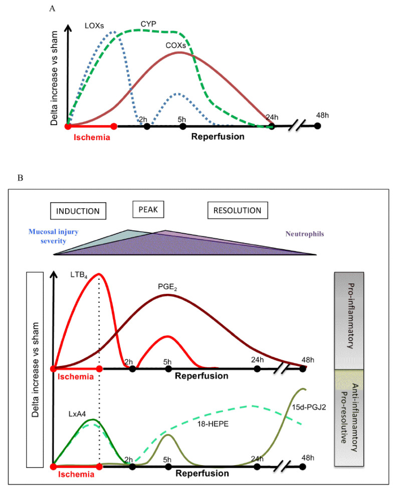
Figure 9.
Changes in the concentrations of various bioactive lipids (specifically products of AA, EPA, and DHA) in ischemia reperfusion injury in mouse intestine. These results are somewhat like those seen in Figure 7 except for the fact that, in this study, LTB4 and cytochrome metabolites of AA/EPA/DHA were also measured. It is possible that different tissues may respond differently to injury but the balance between pro- and anti-inflammatory eicosanoids is somewhat similar. It is noteworthy that PGE2 levels (in conjunction with COXs’ expression) were gradually enhanced from ischemia to reperfusion phase until healing occurs. LXA4 and LTB4 concentrations were elevated during ischemia (up to 2 h) and peaking at 5 h, coinciding with PGE2 levels. These data were taken from ref. [37]. (A) Changes in the activities of COX, LOX and CYP enzymes during ischemia and reperfusion injury. (B) Changes in the concentrations of LTB4, PGE2, LXA4, 18-HEPE and PGJ2 during ischemia and reperfusion injury/inflammation. Notice the relationship between LTB4 and LXA4 (LTB4 > LXA4 and PGE2 > LXA4) during ischemia and reperfusion injury. It may be noted that LTB4 and PGE2 levels are high that. In turn, trigger the production of LXA4 and 18-HETE and PGJ2 that finally induce resolution of inflammation, tissue regeneration and restoration of homeostasis.

