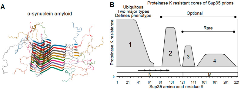
Figure 1.
α-synuclein amyloid and Sup35 prion structure. (A) A typical parallel in-register amyloid structure exemplified by that of α-synuclein (PDB 2N0A) [48]. (B). Map of proteinase K-resistant cores of Sup35 prions. The picture summarizes data for 26 [PSI+] isolates differing in their origin and phenotype [45]. The location of Sup35 N and M domains, as well as of the imperfect oligopeptide repeats, is shown.

