Figure 5.
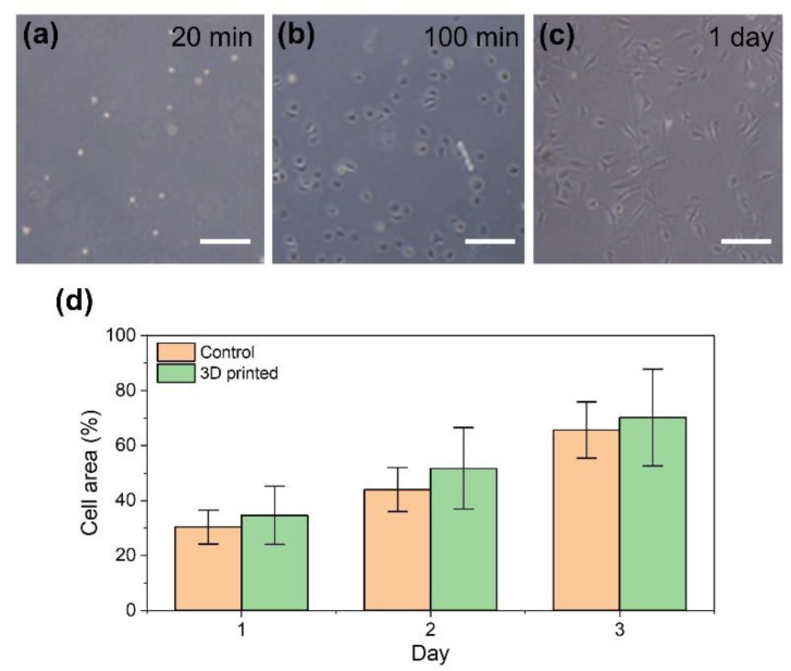
Mouse fibroblast 10T1/2 cells released on a cell culture dish from degraded cell-laden HA-Ph hydrogel by soaking in a medium containing hyaluronidase. Cell morphologies at (a) 20 min, (b) 100 min, and (c) 1 day after treatment for degradation. Growth profiles of the released cells (3D printed) and of those not exposed to 3D printing (control) are expressed in terms of cell area on the cell culture dish. Bars in panels (a–c): 200 µm and panel (d): standard deviation (n = 3).

