Figure 6.
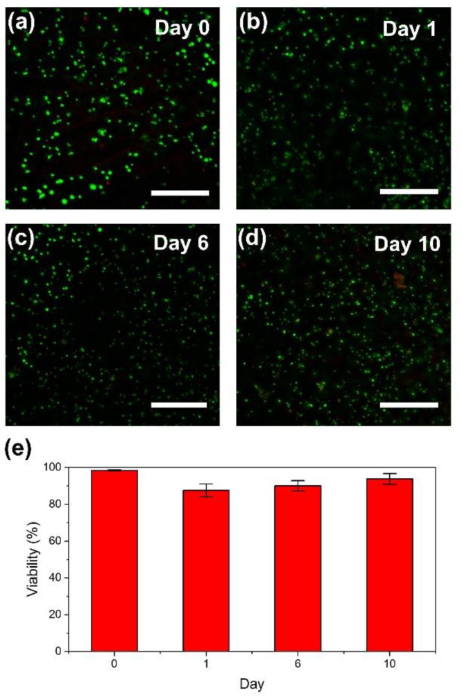
(a–d) Fluorescence microphotographs of mouse fibroblast 10T1/2 cells enclosed in bioprinted HA-Ph hydrogel discs: (a) immediately after printing, at (b) 1 day, (c) 6 days, and (d) 10 days after printing. Live and dead cells showed green fluorescence, attributed to Calcein-AM, and red fluorescence, attributed to PI, respectively. (e) Viabilities of the enclosed cells. Bars in panels (a–d): 200 µm, and in panel (e): standard deviation (n = 3).

