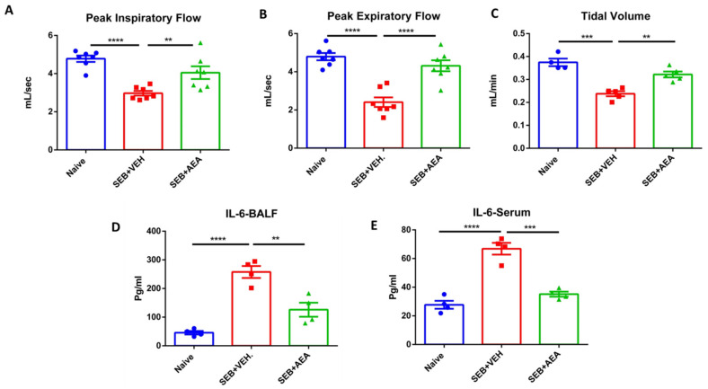
Figure 1.
AEA improves the clinical symptoms of ARDS in the lungs induced by SEB. On day −1, 0, and 1, mice received 40 mg/kg of AEA or VEH I.P. and SEB at a dose of 50 μg/mouse intranasally on day 0. Mice were euthanized 48 h after SEB exposure. The lung functions were assessed using plethysmography. The data shown include clinical functions of the lung, including (A) peak inspiratory flow, (B) peak expiratory flow, and (C) tidal volume (TV). (D) Measurement of cytokines IL6 in the sera and (E) bronchoalveolar lavage fluid (BALF). In panels (A–C), seven mice were used in each group, and in panels (D,E) four mice were used in each group. One-way ANOVA with post hoc Tukey’s test was used to compare the three groups. The data were confirmed in three independent experiments. The vertical bars represent Mean ± SEM and statistical significance was indicated as follows: * p ≤ 0.05, p ** ≤ 0.01, *** p ≤ 0.001, **** p ≤ 0.0001.

