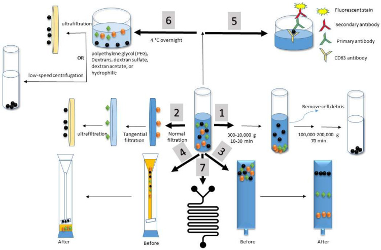
Figure 4.
Isolation techniques for exosomes. The exosomes are represented by the small, black balls. The techniques are represented as separated pathways as follows: (1) ultracentrifugation, (2) ultrafiltration, (3) size exclusion chromatography, (4) hydrostatic filtration dialysis, (5) immunoaffinity, (6) precipitation, and (7) microfluidics.

