Figure 1.
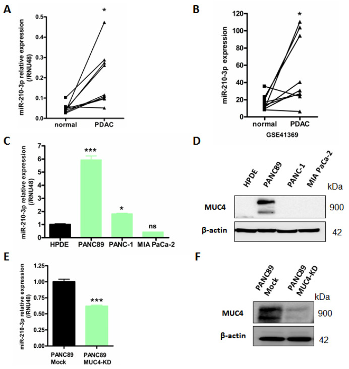
MiR-210-3p is overexpressed in PDAC tissues and pancreatic cancer cell lines compared to normal pancreas. (A) RT–qPCR analysis of miR-210-3p relative expression level in nine paired human pancreatic cancers and their corresponding non-tumoral adjacent tissues. Expression levels are evaluated using 2−ΔCt method (ΔCt = Ct miR-210—Ct RNU48). (B) Analysis of miR-210-3p expression level in GSE41369 PDAC dataset using GEO2R analyzer. (C–E) RT–qPCR analysis of miR-210-3p relative expression in PANC89, PANC-1 and MIA PaCa-2 pancreatic cancer cells, HPDE normal human pancreatic ductal cells (C) and PANC89 Mock and MUC4-KD cells (E). Expressions were determined according to the 2−ΔΔCt method (ΔΔCt = (Ct miR-210—Ct RNU48)—Ct HPDE). Three independent experiments were performed. (D–F) Western blotting analysis of MUC4 and β-actin expression in PANC89, PANC-1, MIA PaCa-2, HPDE (D) and Mock and MUC4-KD PANC89 cells (F). * p < 0.05 and *** p < 0.001 indicate statistical significance compared to normal tissues. ns indicates no statistical significance. At least three independent experiments were conducted.

