Figure 9.

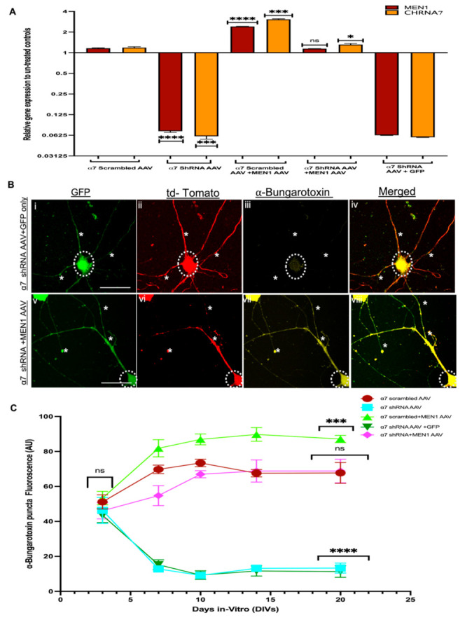
Overexpression of neuron-specific menin restores α7 nAChRs puncta expression in α7 KD hippocampal neurons (in vitro). (A) Summary data, fold change gene expression in from α7 nAChR scrambled AAV, α7 nAChR shRNA AAV, α7 nAChR scrambled AAV+MEN1, α7 nAChR AAV+MEN1AAV and α7 nAChR AAV+GFP AAV hippocampal neuronal cultures relative to untreated controls, determined by qPCR (n = 6, three independent experiments each, DIV 20). Upregulation of MEN1 and CHRNA7 genes was observed in α7 nAChR scrambled AAV+MEN1, whereas the restoration of MEN1 and CHRNA7 was observed in α7 nAChR AAV+MEN1AAV neuronal samples. (B) ICC characterization of α7 KD+MEN1 AAV hippocampal neuronal cultures (Bv–viii), co-transduced with GFP-labelled MEN1 encoding AAV (Bv) compared to scrambled controls (Bi-iv) co-transduced with GFP only AAV (n = 25 images, six independent samples, representative image). tdTomato-positive neurons (Bii,vi) and GFP-positive neurons (Bi,v) labelled with α-bungarotoxin (Biii,vii) and merged (Biii,vi), (Bvii) exhibits significantly increased expression of α7 nAChRs in tdTomato+GFP-positive hippocampal pyramidal neurons compared to α7 KD+GFP only control (Biii). (C) Summary data, normalized bungarotoxin expression in neurons from α7 nAChR scrambled AAV, α7 nAChR shRNA AAV, α7 nAChR scrambled AAV+MEN1, α7 nAChR AAV+MEN1AAV and α7 nAChR AAV+GFP AAV neuronal cultures compared to relevant controls (n ≥ 35 images, ≥7 independent samples) DIV 3, 7, 10, 14 and 20.Scale bars, (Bi–iv) 15 and (Biv–vi) 12 μm. White dotted circles indicate GFP-positive+tdTomato-positive pyramidal neurons. White asterisks on neurites indicate α7 nAChRs expression in tdTomato+ GFP-positive neurites. Statistical significance (one-way ANOVA followed by Tukey’s multiple comparison test) **** p < 0.0001, *** p < 0.001, * p < 0.1, ns p > 0.9999. See Supplementary Tables S15 and S16.
