
Figure 10.
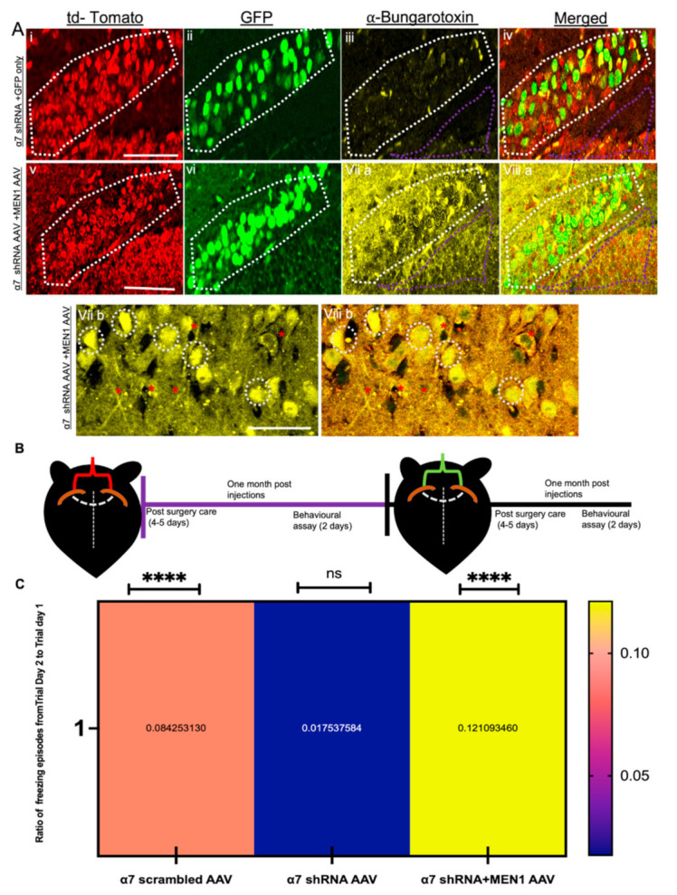
Overexpression of exogenous menin in the a7 nAChRs KD mice rescues hippocampus-dependent learning and memory. (A) IHC characterization of α7 KD+MEN1 AAV hippocampal brain slices (Bv–viii), compared to scrambled controls (Bi–iv) co-transduced with GFP only AAV (n = 25 images, six independent samples, representative image). tdTomato-positive neurons (Bi,v) and GFP positive neurons (Bii,vi) labelled with α-bungarotoxin (Biii,viia,b) and merged (Biv,viiia,b), Bviia,b exhibits significantly increased expression of α7 nAChRs in tdTomato+GFP-positive hippocampal pyramidal neurons compared to the α7 KD+GFP only control (Biii).Panels viib and viiib display magnified images to display increased α7 nAChRs in hippocampal neurons (α7 KD+MEN1 AAV). (B) Illustrative representation of experimental protocol timeline for bilateral stereotaxic injections in CA1 coordinates; red indicates α7 nAChR scrambled AAV, α7 nAChR shRNA AAV injections, green indicates α7 nAChR scrambled AAV+MEN1, α7 nAChR AAV+MEN1AAV and α7 nAChR AAV+GFP AAV viral administration. (C) Heat map for contextual fear conditioning test in three groups, representing the ratio of freezing episodes on day 2 compared to trial day 1. White dotted region indicates GFP-positive+tdTomato-positive pyramidal neurons. White dotted circles show increased expression of α7 nAChRs in α7 KD+MEN1 AAV neurons. Purple dotted shape indicate synaptic puncta region. Red asterisks indicate α7 nAChRs puncta expression in tdTomato+GFP-positive neurites. Statistical significance (one-way ANOVA followed by Tukey’s multiple comparison test) **** p < 0.0001, ns p > 0.05. See Supplementary Tables S17 and S18.