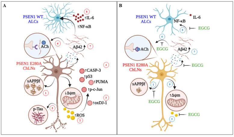
Figure 8.
Schematic representation of the protective effect of EGCG on PSEN 1 E280A ChLNs. (A) Intracellularly accumulation of sAPPβf (step 1) generates H2O2 (s2), which in turn oxidizes the OS sensor protein DJ-1 at Cys106-SH residue into DJ-1Cys106-SO3 (s3) and activates a domino-like, pro-death signaling mechanism (s4) by triggering the activation of transcription factor P53 and c-JUN, BH-3-only protein PUMA and CASP-3. Interestingly, (i)sAPPβf induces the phosphorylation of protein TAU (p-TAU, s5). Besides, PSEN 1 E280A ChLNs do not respond to ACh stimuli i.e., intracellular transient Ca2+ increase is missing due to extracellular interaction between (e)Aβ42 and nicotinic (n)ACh receptors (s6). Therefore, the (i) sAPPβf/Aβ42-induced signaling process (s1–s6) leads ChLNs to structural alterations, cell death (apoptosis), as well as intracellular Ca2+ dysfunction. To aggravate matters, the (e)Aβ42 (s7) induces ALCs to secrete pro-inflammatory cytokine IL-6 through activation of transcription factor NF-κB (s8). (B) Upon exposure to EGCG, mutant ChLNs show normal features such as no oxidized protein DJ-1 (s3), unaltered ∆Ψm (s9), and intact nuclei morphology. Moreover, EGCG partially inhibits the aggregation of (i)sAPPβf (s1) and blocks the generation of H2O2 (s2). As a result, there is no further activation of pro-death proteins (s4), and phosphorylation of protein TAU (s5). Furthermore, EGCG reestablishes the PSEN1 E280A ChLNs response to ACh (s6–s7) and protects ALCs against (e)Aβ42-induced pro-inflammation stimuli (s8).

