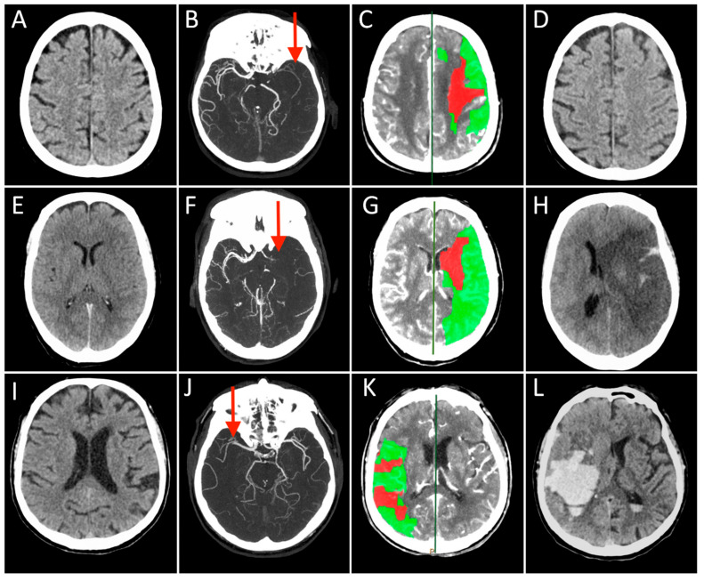
Figure 1.
Each row represents a single patient’s images derived in different modalities and timepoints (Case 1 presented in (A–D), Case 2 in (E–H), and Case 3 in (I–L)). The first three columns on the left show the Non-Contrast-CT (NCCT), multiphase CT Angiography (CTA), and CT Perfusion (CTP) performed at hospital arrival, while the last column on the right displays the NCCT acquired at 24 h after stroke. On presenting NCCT (A,E,I) no early ischemic changes can be seen in the brain tissue in Case 2 and 3 (ASPECT score = 10), while a tissue swelling was detected in the right insular lobe in Case 1 (ASPECT score = 9, not shown). The mCTA images identified proximal (M1 segment) Middle Cerebral Artery (MCA) occlusion in the left hemisphere in (B,F), and contralateral side in (J) (red arrows). CTP showed in all cases a small infarct core corresponding to CBV lesion (red) and a large ischemic penumbra consisting of the difference between MTT and CBV lesions (green), representing the expected “salvageable” tissue after recanalization. In these patients, CBV lesion volume ≤ 50% of MTT lesion size. All patients were treated with combined IVT and MT, obtaining a complete recanalization, but the 24 h NCCT showed three different conditions: no ischemic lesion visible (D), complete MCA territory infarct associated with a massive cerebral oedema (H), and a vast hemorrhagic transformation of the ischemic lesion.

