
Fig 3. CD40L deficiency results in reduced Iba1+ microglial inflammation in the brain.
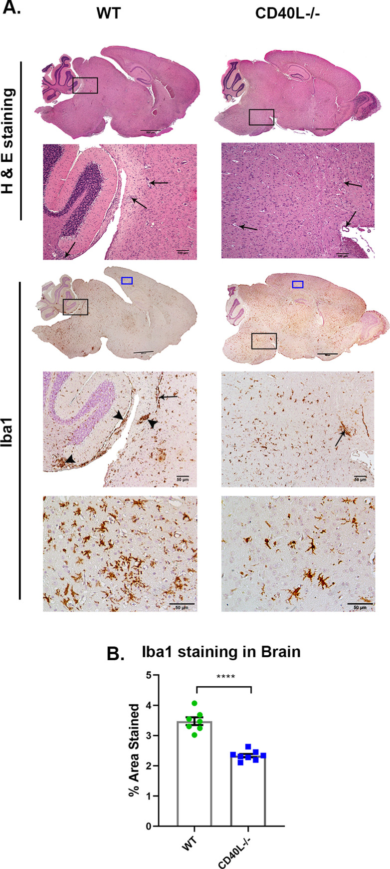
On day 5 p.i., sections of brains (A) from RSA59 infected (25000 PFUs) WT and CD40L-/- mice were stained with H&E and immunohistochemically for Iba1. The boxed areas are shown at higher magnification below the corresponding brain midsagittal sections. Black arrows in the zoomed sections mark characteristic perivascular cuffing, and black arrowheads mark microglial nodule formation. High magnification images corresponding to blue-lined rectangles from the cortical regions show the distinct morphology and distribution of Iba1+ cells. Scale bar, 500μm, 50μm. (B) Shows quantification of Iba1 staining in the brain. Results were expressed as mean ± SEM from 3–4 independent biological experiments (N = 4–5). *Asterisk represents statistical significance calculated using unpaired Student’s t-test and Welch correction, p<0.05 was considered significant. ****p<0.0001.