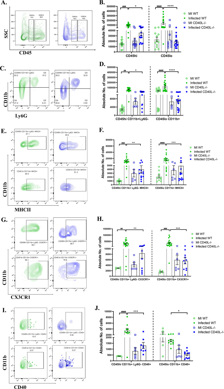
Fig 4. CD40L deficiency causes impaired accumulation and activation of peripheral monocyte/macrophage and the brain resident microglia at the acute phase of inflammation.
On day 5 p.i., brains from MI and RSA59 infected (25000 PFUs) WT and CD40L-/- mice were harvested for flow cytometry analysis and stained for CD45, CD11b, MHCII, CX3CR1, and CD40. The green color denotes WT, and blue indicates CD40L-/- mice. (A) Representative flow cytometry contour plots showing percentages of overall CD45hi and CD45lo cell population after gating on singlets followed by live cells and absolute cell numbers from infected sets are represented in (B) comparing WT and CD40L-/- from MI and infected mice groups. Percentages of CD45 gated cells assessed for CD45hiCD11b+ (peripheral derived monocyte/macrophage) and CD45loCD11b+ (brain resident microglia) from infected sets are presented in flow cytometry plots (C), and absolute numbers of MI and infected sets are graphically represented in (D) comparing WT and CD40L-/- groups. Representative flow cytometry contour plots indicate percentages of MHCII+ (E), CX3CR1(G), and CD40 (I) expressing CD45hiCD11b+ and CD45loCD11b+ cells comparing infected WT and CD40L-/- mice, and the absolute numbers of MI and infected sets are graphically represented in F, H, and J, respectively. Results were expressed as mean ± SEM from 3 independent biological experiments (N = 4). *Asterisk (MI WT v/s infected WT) and #hash (infected WT v/s infected CD40L-/-) represents statistical significance calculated using unpaired Student’s t-test and Welch correction, p<0.05 was considered significant. *p<0.05, **p<0.01, ***p<0.001, ****p<0.0001.

