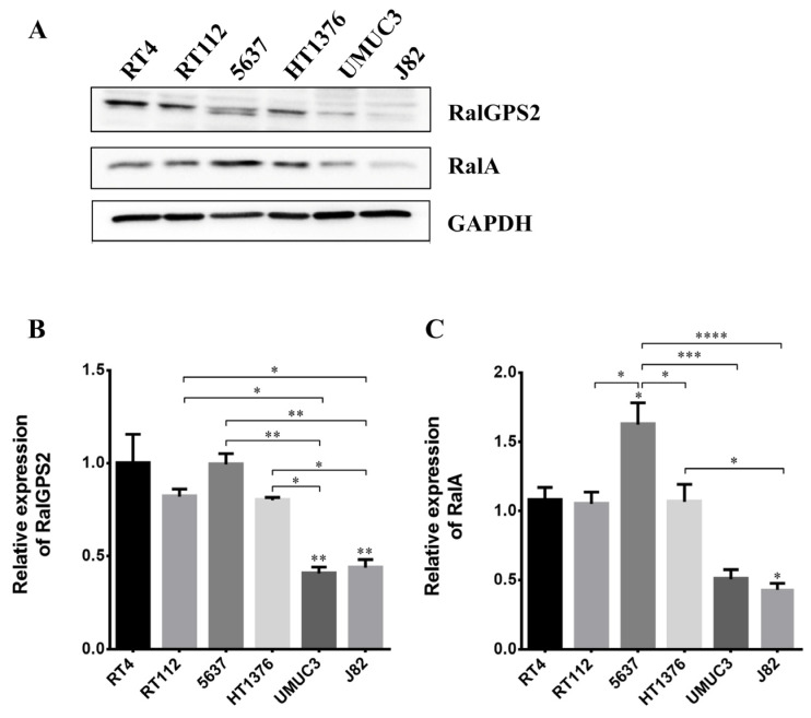
Figure 5.
RalGPS2 and RalA expression in bladder cancer cell lines. RT4, RT112, 5637, HT1376, UMUC-3 and J82 cell lysates were separated on SDS-PAGE and blotted to nitrocellulose membrane; blots were probed with anti-RalA or anti-RalGPS2 or anti-GAPDH antibodies. GAPDH was used as a loading control. Panel (A): representative Western blot results are shown. Panel (B): histograms relative to the quantification of RalGPS2 bands. Panel (C): histograms relative to the quantification of RalA bands. Data are expressed as mean ± S.E.M. from three independent experiments. Differences among groups were analyzed using a one-way analysis of variance (ANOVA) followed by Tukey’s post hoc test. * p < 0.05, ** p < 0.01, *** p < 0.001, **** p < 0.0001.

