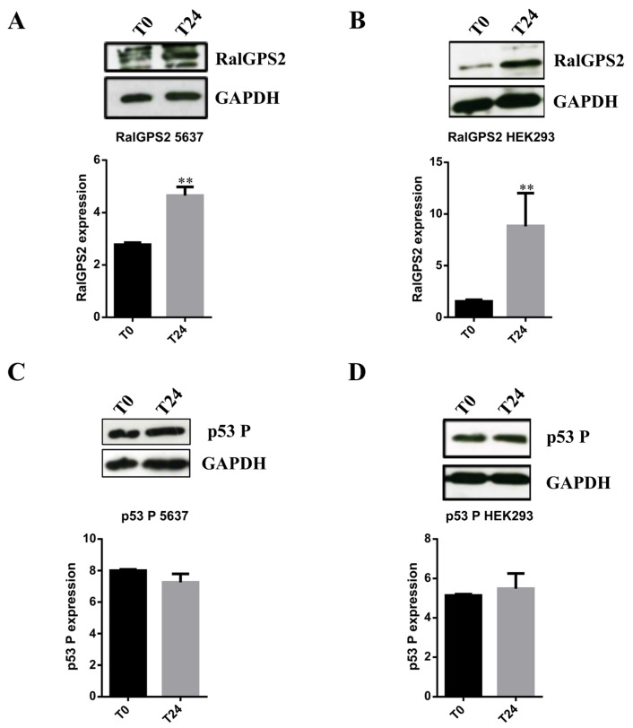
Figure 9.
Stress conditions boost RalGPS2 expression in 5637 and HEK293 cells. 5637 and HEK293 cell lysates were separated on SDS-PAGE and blotted to nitrocellulose membrane; blots were probed with anti-RalGPS2 or anti-phospho p53 (p53 P) or anti-GAPDH antibodies. GAPDH was used to normalize sample loading. Panels (A,C): expression of RalGPS2 and p53 P in 5637 cells treated with LSA medium for 24 h. The panels show representative western blot results and the quantification of (A) RalGPS2 or (C) p53 P expression. Panels (B,D): expression of RalGPS2 and p53 P in HEK293 cells treated with HS+H2O2 medium for 24 h. The panels show representative western blot results and the graphical representation of (B) RalGPS2 or (D) p53 P expression. Data are expressed as mean ± S.E.M. from three independent experiments. Differences among groups were analyzed using Student’s t-test. ** p < 0.01.

