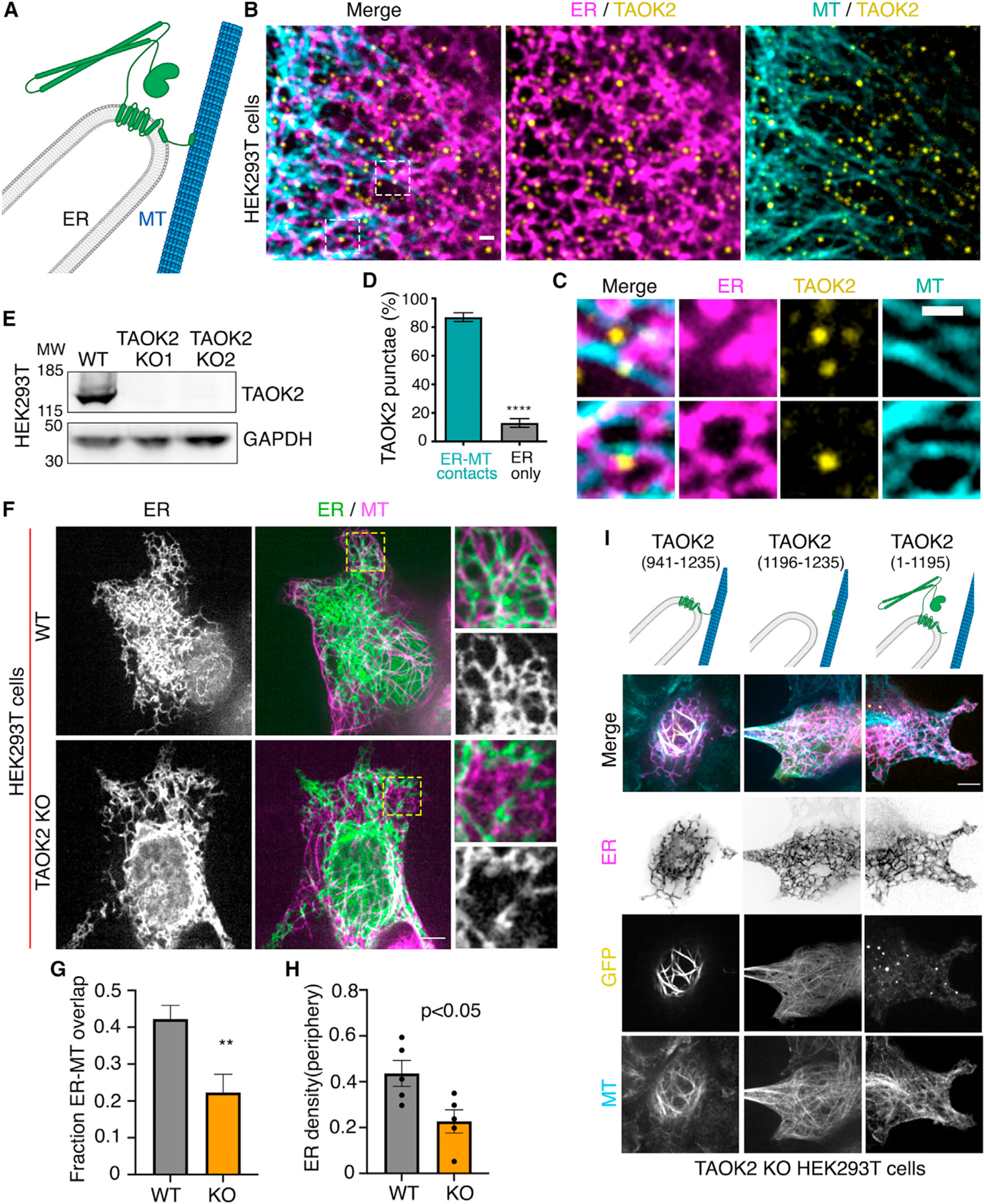
Figure 3. TAOK2 is an ER-MT tethering protein kinase.

(A) Schematic representation of TAOK2 with cytoplasmic-facing kinase domain, membrane bound 6 transmembrane helices and amphipathic region, and C-terminal tail directly bound to MT.
(B) Cells transfected with GFP-TAOK2 (yellow), ER-mRFP (magenta), and MT dye (cyan). Scale bar: 1 μm.
(C) Magnified images displays individual TAOK2 (yellow) punctae on ER tubules in contact with MT (cyan). Scale bar: 1mm.
(D) Percentage of TAOK2 punctae co-localized with ER and MTs in HEK293T cells. Values indicate mean ± SEM; n = 10 cells with at least 50 punctae per cell were analyzed. ****p < 0.0001
(E) Western blot of lysate from WT and TAOK2 KO cell lines generated using CRISPR/Cas9 gene editing.
(F) WT and TAOK2 KO cells transfected with ER-mRFP (green) and mEmerald-ensconsin (magenta) to visualize MTs. Peripheral ER is magnified and shown for both WT and TAOK2 KO cells. Scale bar: 5 μm.
(G) Ratio of peripheral ER area in contact with MT in 100-μm2 region for WT and TAOK2 KO cells. Values indicate mean SEM; n = 6 cells from 3 experiments, t test with Welch’s correction. **p < 0.01
(H) ER density measured in WT and TAOK2 knockout cells. Values indicate mean ± SEM, n = 6 cells from 3 experiments, t test with Welch’s correction.
(I) Topology of TAOK2 deletion constructs. KO cells expressing indicated TAOK2 constructs are shown where TAOK2 (yellow), ER-mRFP (magenta) and MTs (cyan) are shown in merged and grayscale. Scale bar: 5 μm. See also Figure S2.
