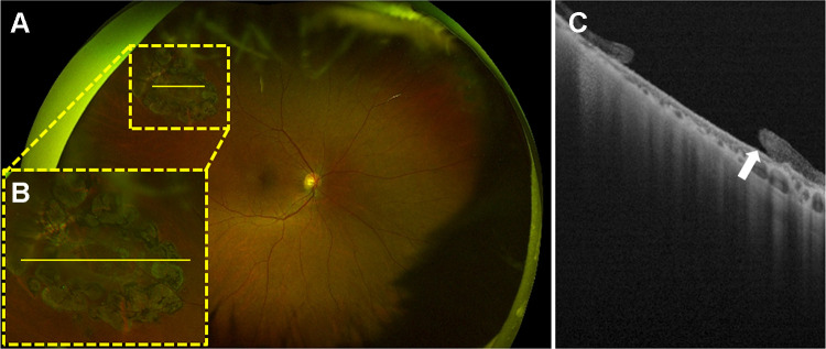
Figure 2.
A multimodal imaging of a 74-year-old female with a peripheral retinal hole treated by a laser photocoagulation in the right eye. (A) An ultra-widefield fundus photograph. A retinal hole surrounded by laser scars is observed in the mid-peripheral area (yellow-dotted square). (B) A magnified image of the yellow-dotted square on (A). (C) An ultra-widefield swept-source optical coherence tomography image of the yellow line (A and B). The edge of the retinal hole is slightly detached (white arrow).

