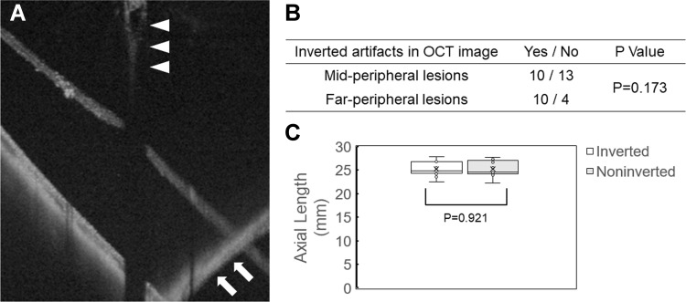
Figure 4.
An inverted optical coherence tomography image. (A) Swept-source optical coherence tomography (OCT) image of the peripheral retinal tear. A vitreoretinal traction is observed (white arrowheads). The OCT image is inverted (white arrows). (B) Inverted artifacts in OCT image related to the location of the retinal degenerations. There was no significant difference between the mid-periphery and far-periphery (P=0.173). (C) The mean axial length of the inverted and noninverted group. There was no significant difference (P=0.921).

