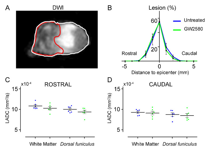
Figure 2.
Long-term GW2580 treatment has no effect on lesion extension and longitudinal diffusivities. Representative ex vivo DW-MRI located at the lesion epicenter 6 weeks after injury (A). Axial spinal cord slice is delineated in white and lesion (here at the epicenter) in red. Line curves showing lesion percentages at epicenter, lesion extensions, and lesion volumes (area under the curve) in untreated (blue) and GW2580-treated (green) groups (B). Longitudinal apparent coefficient diffusion (LADC) rostral (C) and caudal (D) to the lesion in the white matter (excluding the dorsal funiculus) and the dorsal funiculus. Results are expressed as mean ± SEM per time-point in untreated (blue) and GW2580-treated groups (green). Each dot corresponds to a minimum of 8 sections (1 mm interval between each section). Statistics: Student’s unpaired t-test. Number of animals: 6 mice were used per experiment and per condition.

