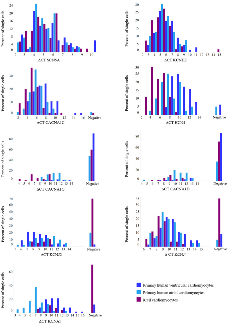
Figure 1.
Histograms of expression parameters. Comparisons of distributions of single-cell expression data (ΔCT) between atrial and ventricular human primary cardiomyocytes and iCell cardiomyocytes. For total n see Table 3. While for some ion channels (SCN5A, KCNH2, CACNA1C, and KCNJ4) the distributions between primary human atrial and ventricular cardiomyocytes are comparable, the distributions of HCN4, CACNA1D, CACNA1G, and KCNA5 are left-shifted (higher expression) in primary human atrial cardiomyocytes compared to primary human ventricular cardiomyocytes. With respect to KCNJ2, distribution is left-shifted (higher expression) for human primary ventricular cardiomyocytes compared to their atrial counterpart. Comparing distributions between the two primary cardiomyocyte groups and iCell cardiomyocytes shows a shift to the left (higher expression) for the pacemaking-associated ion channels HCN4, CACNA1G, and CACNA1D in iCell cardiomyocytes.

