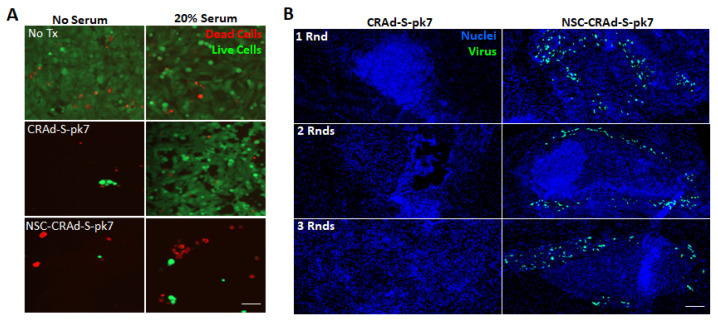
Figure 2.
NSCs protect CRAd-S-pk7. (A) Representative fluorescence images of day 7 U251 brain cancer cell cultures stained with calcein-AM and ethidium bromide to visualize live (green) and dead (red) cells, respectively. Cultures were treated with either free CRAd-S-pk7 or dose-matched NSC-Crad-s-pk7 with and without the addition of 20% human serum. Scale bar = 50 µm and applies to all images. (B) Immunocompetent C57/BL-6 mice (8 weeks old females) bearing 4-day old intracranial GL261 glioma (2 × 10³ cells) received either 1, 2, or 3 weekly rounds of intra-tumoral CRAd-S-pk7 (2.5 × 10⁷ IU) or dose-matched NSC-CRAd-S-pk7. Brains were harvested, fixed, and cryosectioned 1 day after treatment. Brain slices were stained with anti-hexon FITC to visualize CRAd-S-pk7, and nuclei counterstained with DAPI. Scale bar = 100 µm and applies to all images.

