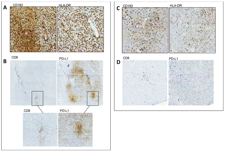
Figure 1.
IHC performed on FFPE specimens of Pt#1 and Pt#3. In IDH1 wt rGBM (Pt#1), (A) adjacent representative sections show a high frequency of GAMs expressing CD163 (left) and HLA-DR(right). (B) the distribution of CD8+ T cells (left) is clustered near the blood vessels and within the tumor mass around the tumor cells, and the same areas of the adjacent sections express high levels of PD-L1. The black rectangle identifies intensely infiltrated CD8+ T cells and the corresponding area where tumor cells overexpress PD-L1. In IDH1-mutant (mut) rGBM (Pt#3), (C) adjacent representative sections show low frequency of GAMs expressing CD163 (left) and HLA-DR (right), with a prevalence of small size cells. (D) CD8+ TILs (left) are rare, and, when present, the distribution is scattered. PD-L-1 (right) expression is not detected.

