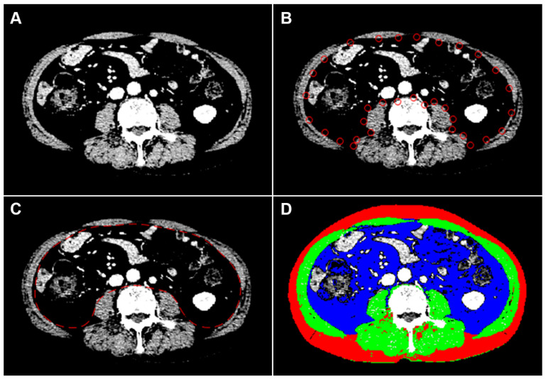
Figure 1.
An example of semiautomatic quantification of body composition in a 75-year-old man with metastatic hormone-sensitive prostate cancer. To highlight the muscle boundary, the intensity of the CT image is linearly transformed into 0 to +100 HU (A). After semiautomatic manipulation (B), the boundary between the muscles and the inner tissues is detected using the active contour method by minimizing a cost function, dividing CT images into inner and outer regions (C). Pixels in the fat and muscle are then identified using cut-off values of −300 to −50 HU and −29 to +150 HU, respectively (D). The cross-sectional areas of muscle, subcutaneous fat, and visceral fat are measured to be 140.03 cm2, 118.69 cm2, and 181.37 cm2, respectively. Green-colored, red-colored, and blue-colored areas represent muscle, subcutaneous fat, and visceral fat, respectively; CT, computed tomography; HU, Hounsfield unit.

