Figure 1.
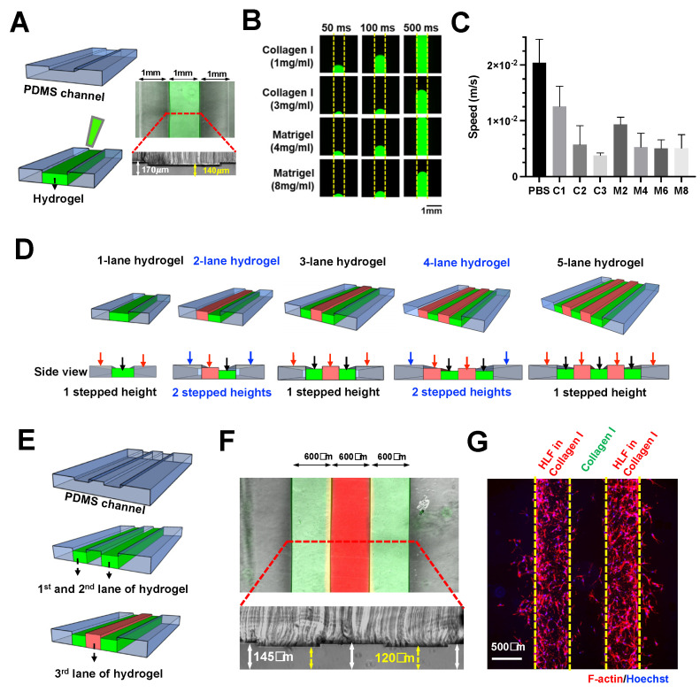
Scalable hydrogel patterning in enclosed microchannel using stepped height features. (A) Schematic illustration of stepped height-based hydrogel patterning in a single lane hydrogel chip. Overlaid fluorescence and brightfield image of the chip loaded with FITC-laden Collagen I (3 mg/mL) in the hydrogel channel (top). Cross-sectional view of the chip, white arrow indicates channel height of 170 μm, while yellow arrows indicate channel height of 140 μm (bottom). (B) Fluorescence images showing the gel loading process at different timepoints for Collagen I (1, 3 mg/mL, FITC-laden) and Matrigel (4, 8 mg/mL, FITC-laden). Yellow dotted line indicates the channel boundary. (C) Loading speed of 1 PBS, Collagen I (1, 2, and 3 mg/mL), Matrigel (2, 4, 6, and 8 mg/mL) in the one-lane hydrogel chip. (D) Schematic illustrating the concept of multi-lane hydrogel confinement using a single stepped height for odd number of lanes, and two stepped heights for even number of lanes. Black arrows indicate the first-layer channels, red arrows indicate the second-layer channels, and blue arrows indicate the third layer channels. (E) Schematic of sequential three-lane hydrogel loading. (F) Overlaid fluorescence and brightfield image of the chip loaded with FITC-laden Collagen I (3 mg/mL) in the first and third lanes, and R6G-laden Collagen I in the second lane (middle). Cross-sectional view of the chip, white arrow indicates channel height of 145 μm, while yellow arrows indicate channel height of 120 μm (bottom). (G) Fluorescence image of the chip containing two lanes of HLF-laden Collagen I with a cell-free Collagen I in between. (F-actin–red, Hoechst–blue).

