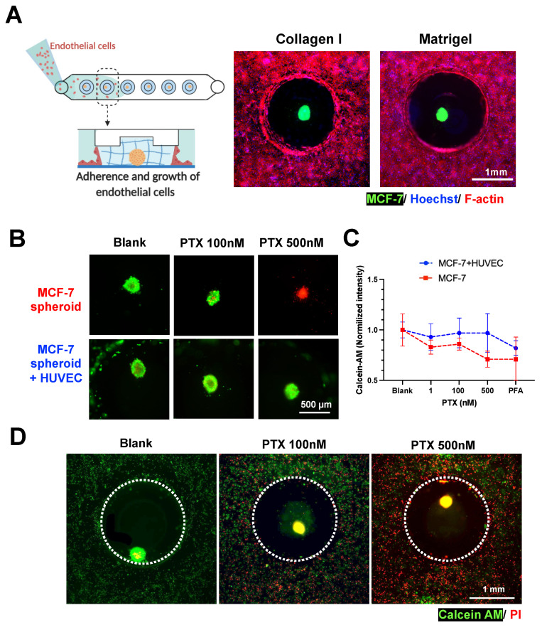
Figure 5.
Spheroid-in-gel formation and co-culture with endothelial cells. (A) Co-culture of MCF-7 spheroids with endothelial cells (HUVEC) in collagen and Matrigel. MCF-7 was labelled with DiO, F-actin—red, and Hoechst—blue. Schematics were created with BioRender.com. (B) Merged fluorescent images illustrating viability of spheroid after PTX treatment. (Calcein-AM—green and PI—red) (C) Normalized fluorescence intensity of Calcein-AM for spheroids with or without HUVEC co-culture after 3 days of treatment with PTX 1, 100, and 500 nM. A PFA-fixed spheroid was used as negative control. Data are presented as the mean SD. (n = 3) (D) Merged fluorescent images illustrating viability of spheroids and HUVEC in co-culture after 3 days of PTX treatment. (Calcein-AM—green and PI—red).

