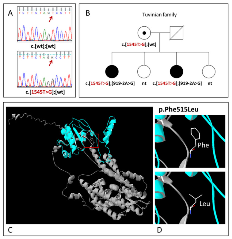
Figure 2.
(A) Identification of variant c.1545T>G (p.Phe515Leu) by Sanger sequencing; (B) The pedigree of the Tuvinian family demonstrating the segregation of variant c.1545T>G (p.Phe515Leu) in compound with recessive mutation c.919-2A>G with HL. Deaf individuals are shown by black symbols; the variant c.1545T>G (p.Phe515Leu) is shown by red; nt—not tested; wt—wild-type; (C) The 3D structure of the pendrin protein with localization of variant p.Phe515Leu; (D) Close-up views of wild (Phe515) and mutant (Leu515) types of pendrin.

