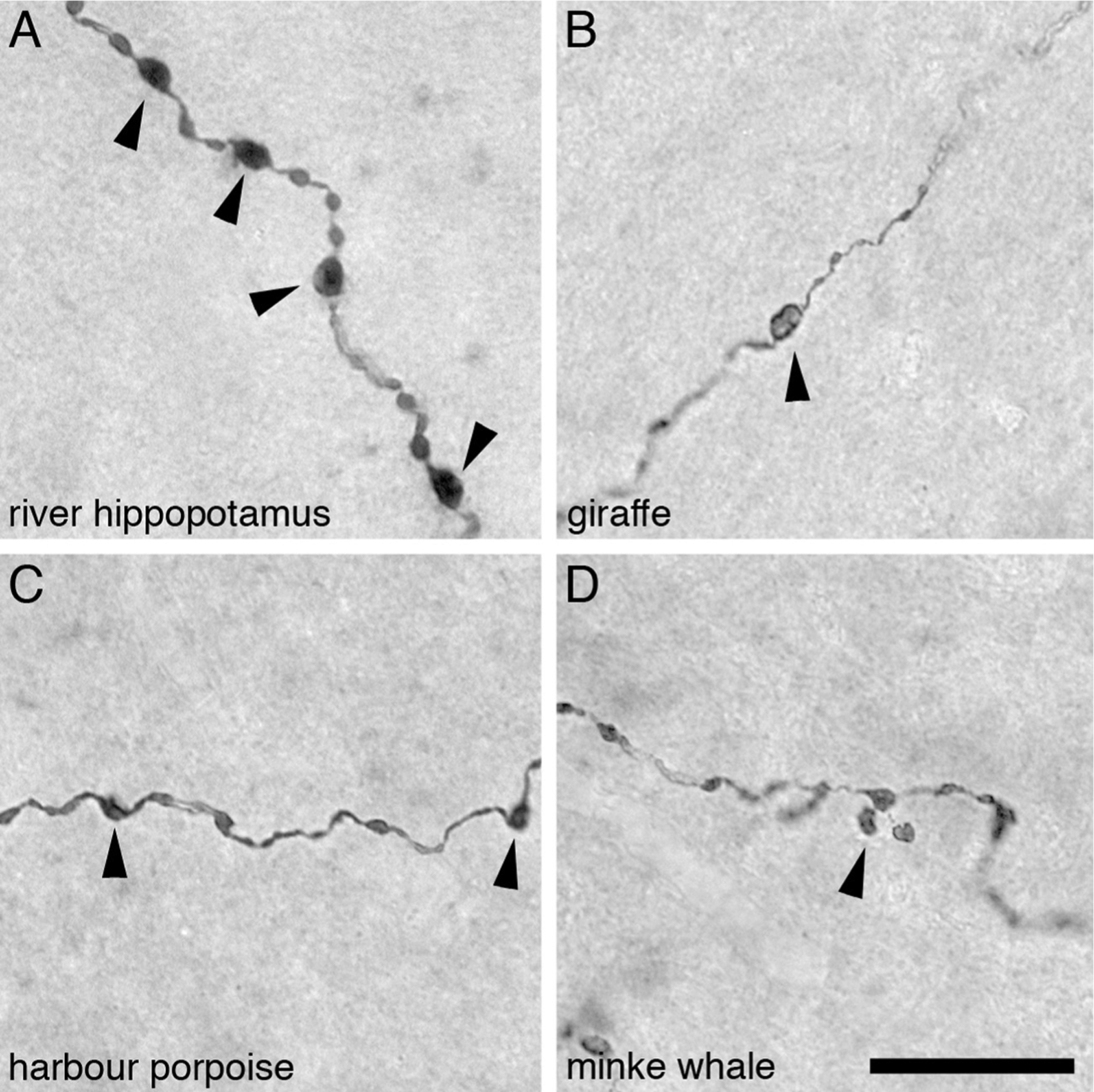
Fig. 4.

High power photomicrographs of orexinergic axons, showing the appearance of the large and small orexinergic boutons, in layer III of the cerebral cortex of four different species of Cetartiodactyla examined in the present study. (A) River hippopotamus (Hippopotamus amphibius). (B) Giraffe (Giraffa camelopardalis). (C) Harbour porpoise (Phocoena phocoena). (D) Minke whale (Balaenoptera acutorostratus). In all images the arrowheads indicate the boutons considered to be large boutons, while the small boutons have been left unmarked. Scale bar in D = 20 μm and applies to all.
