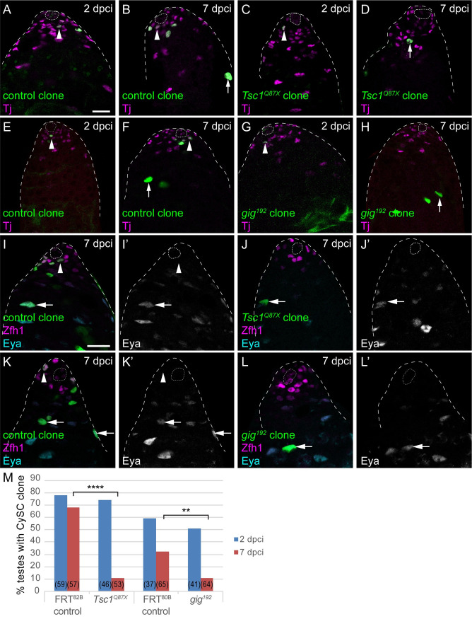
Fig 2. Clonal gain-of-function in Tor activity is sufficient to induce CySCs to differentiate.
A-H. Positively-labelled CySC clones generated by MARCM. Control clones generated with FRT82B (A,B) or FRT80B (E,F) were observed at 2 days post clone induction (dpci) and at 7 dpci to assess clone induction and maintenance rates, respectively. CySCs (arrowheads) were identified as cells expressing Tj (magenta) and in the first row from the hub, differentiated cyst cells (arrows) have larger nuclei and are found away from the hub. Tsc1 mutant (C,D) or gig (Tsc2, G,H) CySC clones could be induced and were present at 2 dpci (C,G) but were rarely observed by 7 dpci (D,H), where differentiated cells were labelled. I-L. Clones (green) in testes labelled with Zfh1 (magenta) and Eya (cyan, single channels in I’-L’). In controls at 7 dpci (I,K), clones contained both Zfh1-expressing CySCs (arrowheads) and Eya-positive cyst cells (arrows). However, Tsc1 mutant (J) or gig mutant (L) clones had differentiated and were composed of Eya-expressing differentiated cells. The hub is outlined with a dotted line. Scale bar in panel A panels represents 20 μm for A-H and scale bar in panel I panels represents 20 μm for I-L. M. Graph showing the percentage of testes containing a CySC clone at 2 (blue) and 7 (red) dpci. The number of testes examined is showed in parentheses for each column. Statistical significance was determined using Fisher’s exact test, **** denotes P<0.0001, ** denotes P<0.005.

