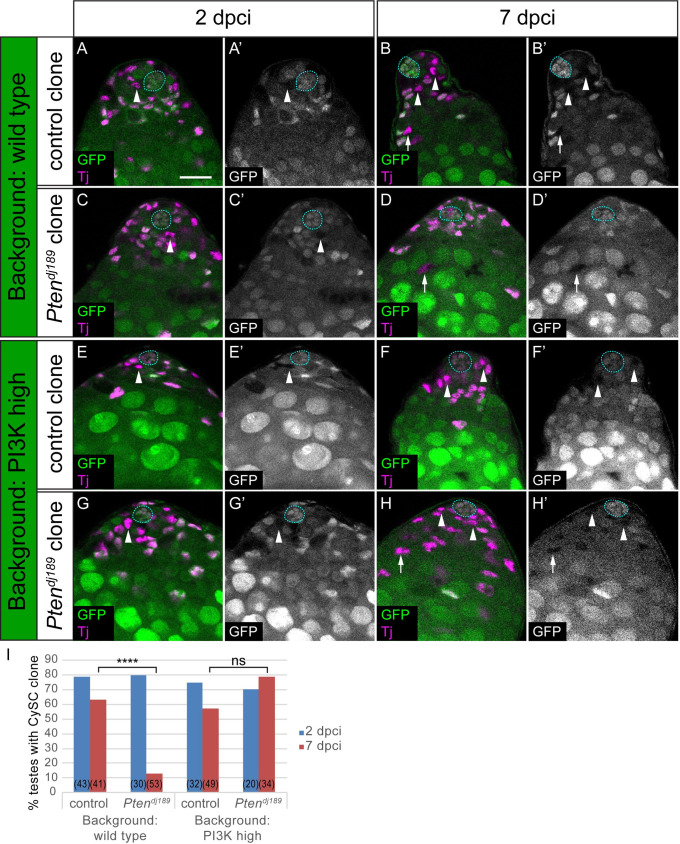
Fig 4. Differentiation of PI3K-high CySCs depends on signalling levels in neighbouring cells.
Control (A,B,E,F) and Pten mutant (C,D,G,H) clones induced by mitotic recombination and marked by loss of GFP expression, generated either in a control background (C587-Gal4>+, A-D) or in a background with elevated PI3K activity (C587-Gal4>Dp110, E-H). CySCs (indicated with arrowheads) were identified as Tj-positive (magenta) nuclei one cell diameter from the hub (outlined with a blue dotted line). While Pten mutant CySC clones were rarely recovered at 7 dpci, mutant cyst cells were observed (D, arrow) indicating differentiation of the clones. By contrast, Pten mutants self-renewed and CySC clones were recovered when PI3K activity was elevated (H, arrowheads). Scale bar in all panels represents 20 μm. I. Graph showing the percentage of testes containing a CySC clone at 2 (blue) and 7 (red) dpci. The number of testes examined is showed in parentheses for each column. Statistical significance was determined using Fisher’s exact test, **** denotes P<0.0001, ns: not significant.

