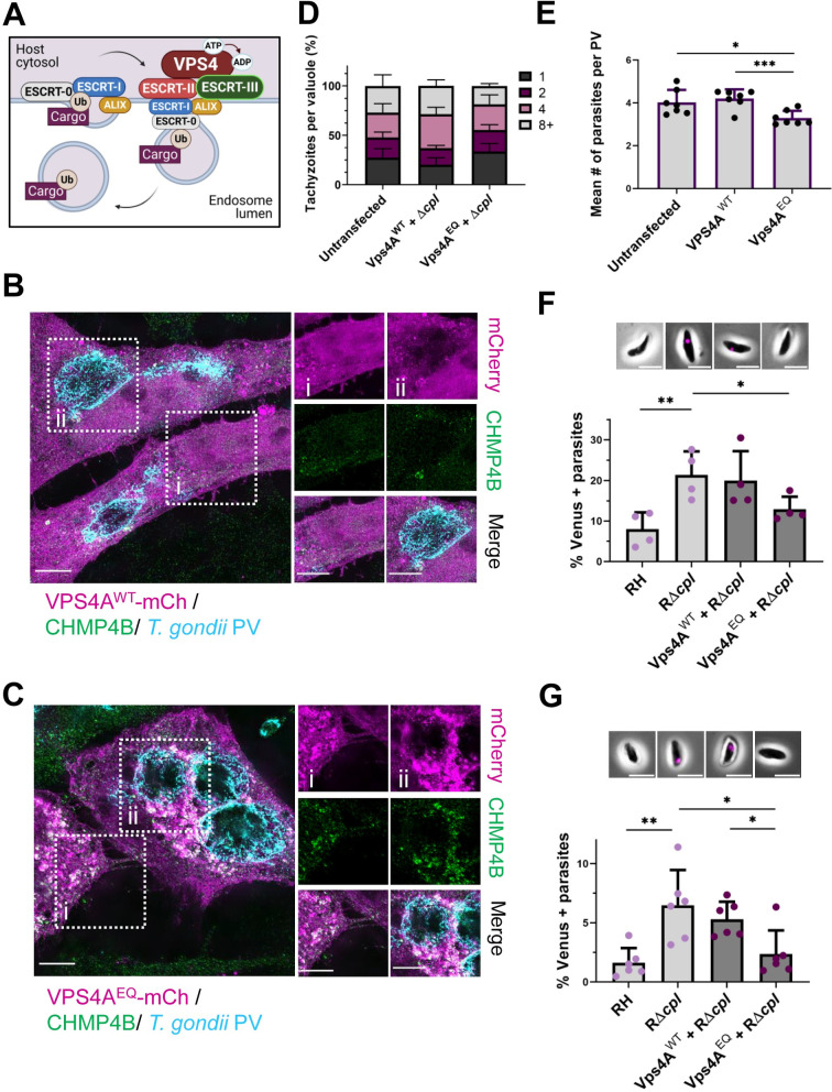
Fig 1. Disruption of the host ESCRT machinery impairs uptake of host cytosolic proteins by Toxoplasma gondii.
A. Schematic of the ESCRT machinery function in MVB formation. Ubiquitinated cargo targeted for degradation is recognized by ESCRT-0 and ECSRT-I, which further recruits ESCRT-II and the ESCRT accessory protein ALIX. At the last steps, ESCRT-III forms spirals at the membrane to mediate membrane constriction and the VPS4 complex facilitates scission and disassembly of the machinery by hydrolyzing ATP. B. Distribution of the ESCRT-III component CHMP4B at the host cytosol (i) and the PV (ii) in HeLa cells transiently transfected with VPS4AWT-mCherry and infected with RH parasites for 24 h. C. Distribution of the ESCRT-III component CHMP4B at the host cytosol (i) and the PV (ii) in HeLa cells transiently transfected with VPS4AEQ-mCherry and infected with RH parasites for 24 h. The PV was labeled with anti-TgGRA7. Images were analyzed by structural illuminated microscopy (SIM). Scale bar is 5 μm. D. Growth assay of RH parasites in HeLa cells transiently transfected with exogenous VPS4WT or VPS4AEQ compared to untransfected control. The percentage of PV with 1, 2, 4 or 8+ parasites was calculated for each cell subset. At least 20 PV were counted per blinded sample. Data represents means from 7 biological replicates. E. Mean number of parasites per PV from growth assay. Statistical analysis was by Student’s t-test. F. Quantification of ingestion of host cytosolic Venus at 30 mpi in RH or RΔcpl parasites harvest from cells were transiently co-transfected with a plasmid encoding cytosolic Venus fluorescent protein expression and exogenous expression of either VPS4AWT or VPS4ADN. Representative images for parasites with ingested host-derived cytosolic Venus (shown in magenta) at 30 mpi included at the top. Scale bar is 5 μm. G. Quantification of host cytosolic Venus ingestion at 24 hpi in RH or RΔcpl parasites. At least 200 parasites were analyzed per blinded sample. Representative images for parasites with ingested host-derived cytosolic Venus (magenta) at 24 hpi are included at the top. Scale bar is 5 μm. Data represents the mean from ≥ 3 biological replicates. Statistical analysis was by Student’s t-test. Only statistical differences are shown. *p<0.05, **p<0.01.

