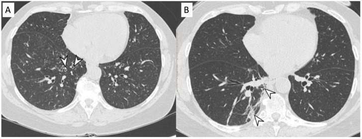
Figure 4.
Microscopic Polyangiitis (MPA). Axial CT scan shows micronodules with centrilobular distribution and diffuse bronchial wall thickening (white arrowheads in (A)). After treatment, 2 years later, axial CT scan shows the appearance of (focal organizing pattern) focal consolidation (white arrowheads in (B)), as clearly depicted in the right lower lobe.

