Figure 7.
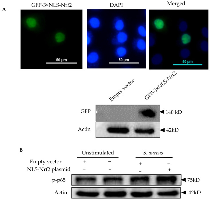
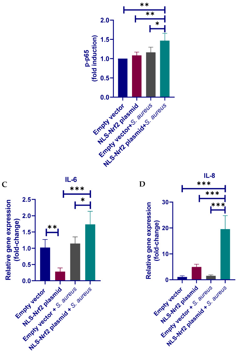
Evaluation of Nrf2 overexpression on the inflammatory responses of pbMECs to S. aureus. (A) Confirmation of Nrf2 overexpression. Cells were transfected with a GFP-expressing Nrf2 plasmid containing tripartite nuclear localization signal (NLS-Nrf2) for 24 h. GFP fluorescence was determined under a microscope and whole-cell extracts were analyzed by immunoblotting with anti-GFP. (B) Evaluation of the effect of Nrf2 overexpression on NF-κB p65 activation in response to S. aureus. The cells transfected with GFP-expressing Nrf2 plasmid were incubated with S. aureus for 6 h. Whole-cell extracts were analyzed by immunoblotting with anti-phospho NF-κB p65 antibody (p-p65). β-actin was used for the equal loading control. p-p65 levels were determined with densitometry analyses after normalization to Actin. Bars are means ± s.d. of three triplicates and are representative of 3 separate experiments. (C,D) Evaluation of the effect of Nrf2 overexpression on the expression of pro-inflammatory cytokines was induced by S. aureus. The cells transfected with GFP-expressing Nrf2 plasmid were incubated with S. aureus for 6 h. Total RNAs were prepared and subjected to qPCR analyses for the mRNA levels of IL-6 and IL-8. The results are the mean ± s.d. of three replicates and are representative of 3 separate experiments (D). * p < 0.05, ** p < 0.01 and *** p < 0.001.


