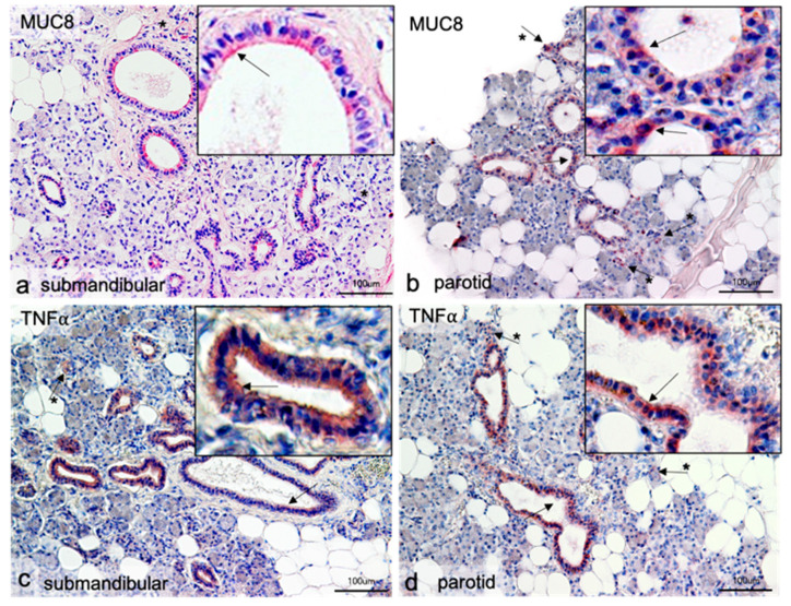
Figure 1.
Immunohistochemical localization of MUC8 (a,b) and TNFα (c,d) in submandibular and parotid glands. In all gland sections, positive antibody responses are visible in the epithelial cells of the intralobular excretory ducts (arrows). The antibody against MUC8 reacts in the submandibular gland at the luminal cell pole of the epithelial cells (insert a); in the parotid, gland single more intense reactions around cell nuclei are visible (insert in b). The antibody reactions against TNFα give similar pictures. Here, more intense signals around cell nuclei are also visible in the Gld. parotidea (insert d). In the Gld. submandibularis, no clear polarization can be observed for TNFα; mostly the whole cytoplasm reacts homogeneously, but there are also single cells with more intense reactions (insert c). Furthermore, single positive cells are seen in the glandular parenchyma in all tissues (arrows with asterisk). Neither serous nor mucosal acini show positive antibody responses.

