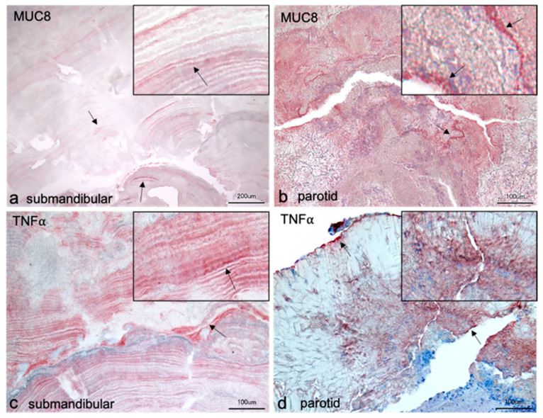
Figure 2.
Immunohistochemical localization of MUC8 (a,b) and TNFα (c,d) in salivary stones from the excretory ducts of the submandibular and parotid glands. The sections of the submandibular stones show layered arrangements. The structure in a is comparable to the switching lamellae of bone; different areas show radially arranged layers around supposedly their own centers. In contrast, the structure in c is reminiscent of the annual rings of trees, as here the layers appear to be arranged around a single center. The antibody against MUC8 reacts positively in individual layers, but alternately shows no signals in other layers. In layers with stronger reactions, punctate granule-like signals are observable (arrows and insert in a). These granule-like signals in the layers are also visible with the antibody against TNFα (arrows and insert in c). In the lower third of the image, a slight increase in reactivity of the red-stained layers is visible in c from bottom to top until a band of blue-stained cell nuclei is seen. No positive signals for TNFα occur in the region of the cell nuclei. In the remaining part of the stone, very distinct layers are seen interspersed with cloud-like amorphous parts. Positive MUC8 reactions occur almost throughout the section of the parotid stone (b). A band of stronger reactivity can be seen, containing cell nuclei (arrows and insert in b). The positive MUC8 signals appear as cloudy shading. Areas with crystalline structure do not show positive responses. The parotid stone in d shows predominantly crystalline structures, which also do not give positive signals. Intense bands at the stone edge and within the stone with intercalated cell nuclei are visible (arrows in d).

